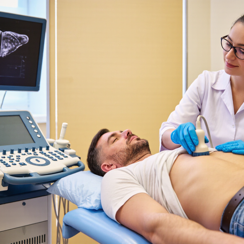
Hepatocellular carcinoma often develops silently, especially in people with cirrhosis, chronic hepatitis B, or chronic hepatitis C. Ultrasound can provide an accessible first look at the liver, but it is not a magic window. Image quality changes with obesity, bowel gas, fatty liver, and operator experience. Small lesions may hide near the diaphragm or blend into a nodular liver background. That limitation deserves honesty.
Dr. Richard G. Barr, a recognized ultrasound specialist and liver-imaging researcher, has emphasized, “The quality of an ultrasound examination depends on the operator.” His point is practical. A reliable Hepatocellular Carcinoma Ultrasound examination requires patient positioning, suitable equipment, careful scanning, and complete documentation. The sonographer should examine every liver segment, compare suspicious areas with surrounding tissue, and record lesion size, margins, echogenicity, vascularity, and location. Doppler findings may add useful information, yet they cannot independently confirm cancer.
This guide explains how clinicians approach suspicious ultrasound findings without overstating what the test can prove. A new mass may appear hypoechoic, hyperechoic, mixed, or nearly invisible. Appearance varies. That is the difficult part. When findings remain uncertain, contrast-enhanced ultrasound, multiphase CT, MRI, laboratory data, or biopsy may be considered by qualified professionals. Screening intervals also depend on individual risk and current clinical guidance. Readers should treat ultrasound as an important detection tool, not a final diagnosis. Careful interpretation protects patients from both missed tumors and unnecessary alarm.
Hepatocellular carcinoma (HCC) is the most common primary liver cancer. It often develops in cirrhosis or chronic hepatitis B and C. IARC’s GLOBOCAN 2022 estimated 866,136 new liver cancer cases worldwide and 758,725 deaths. HCC represents about 75–85% of primary liver cancers, according to global clinical reviews. Symptoms may remain absent during early disease.
Ultrasound is mainly a surveillance tool for people at increased risk. The American Association for the Study of Liver Diseases recommends surveillance every six months, usually with ultrasound and optional alpha-fetoprotein testing. During an examination, clinicians assess liver texture, surface changes, blood vessels, and focal nodules. A suspicious lesion may appear unusually dark, bright, irregular, or vascular. These findings are not diagnostic alone.
If ultrasound detects a nodule measuring at least one centimetre, guidelines generally recommend multiphasic CT or MRI for confirmation. Contrast-enhanced ultrasound can assist selected patients when other imaging is unsuitable. AASLD guidance reports ultrasound sensitivity of roughly 60–80% for liver cancer surveillance, with higher specificity. Early tumors remain easier to miss. Obesity, fatty liver, rib shadows, and operator experience can reduce visibility. That limitation matters. Real-world scanning is less tidy than guidelines suggest. A normal examination lowers concern, but it cannot eliminate risk.
Ultrasound is commonly used for liver cancer surveillance in people at increased risk, especially those with cirrhosis or chronic hepatitis. It is noninvasive and widely available, but its ability to detect very small tumors can be limited.
The ranges show approximate sensitivity reported in clinical studies: ultrasound detects about 60–80% of hepatocellular carcinomas overall, while sensitivity for early-stage disease is commonly lower, around 40–60%. A suspicious or limited examination should be followed by contrast-enhanced imaging and clinical evaluation.
A liver ultrasound can reveal suspicious nodules, altered blood flow, or an uneven liver surface. The examination is painless and usually takes 15 to 30 minutes. The American Association for the Study of Liver Diseases reports that surveillance ultrasound has over 90% specificity, but its sensitivity may be limited, especially for early tumors. Good preparation can improve visibility, though it cannot guarantee a clear scan.
Most hospitals request fasting for six to eight hours before the appointment. Fasting reduces stomach and bowel gas, which can obscure the liver, and keeps the gallbladder distended. Small sips of water are often allowed. Avoid chewing gum, smoking, and carbonated drinks during this period. Wear loose clothing so the upper abdomen can be exposed easily. Do not stop prescribed medicines unless the imaging center or clinician advises it.
Bring previous scans, liver-function results, and a list of medicines. Tell the sonographer about cirrhosis, hepatitis, abdominal surgery, or recent pain. The European Association for the Study of the Liver supports ultrasound surveillance at six-month intervals for people at high risk. However, obesity, fatty liver, and bowel gas may weaken image quality. In such cases, the report may recommend contrast imaging or MRI. Preparation is helpful, not perfect. Follow the local instructions, even when they differ slightly.
Ultrasound can reveal suspicious liver lesions before symptoms appear. During a scan, the clinician studies the liver’s texture, shape, and blood flow. A concerning nodule may look darker or brighter than nearby tissue. It can also have uneven margins, internal vessels, or a distorted surrounding liver pattern. Small details matter.
Color Doppler may show increased arterial blood flow inside a lesion. This finding raises concern, but it does not prove hepatocellular carcinoma. In experienced hands, contrast-enhanced ultrasound can provide more clues. Rapid enhancement during the arterial phase, followed by washout, may suggest malignant behavior. The timing and strength of these changes require careful interpretation.
Ultrasound is not flawless. A fatty liver can hide small nodules, while ribs, bowel gas, or poor breath-holding may reduce image quality. Some harmless lesions can also look alarming. That uncertainty matters. Radiologists compare current images with earlier examinations and consider cirrhosis, hepatitis, blood tests, and symptoms. When findings remain suspicious, multiphasic CT, MRI, or tissue sampling may be recommended according to clinical guidelines. A careful report should describe the lesion’s size, location, appearance, vascular pattern, and technical limitations. One overlooked limitation can change the next clinical decision.
| Ultrasound Dimension | Typical Observation | Why It May Raise Suspicion | Recommended Clinical Response |
|---|---|---|---|
| Patient risk profile | Cirrhosis, chronic hepatitis B, chronic hepatitis C, or other chronic liver disease | Hepatocellular carcinoma is more likely in patients with established chronic liver disease, particularly cirrhosis. | Use a structured surveillance program when clinically appropriate. Ultrasound surveillance is commonly performed every 6 months in at-risk adults. |
| Focal liver lesion | A new, enlarging, or previously undocumented nodule or mass | A focal observation may represent HCC, but benign lesions and other malignancies can have similar appearances on conventional ultrasound. | Document location and maximal diameter, compare with prior studies, and arrange diagnostic multiphasic CT or MRI when indicated. |
| Lesion size | Subcentimeter observation or lesion measuring at least 1 cm | Size influences the next step, although size alone cannot diagnose HCC. | For an observation smaller than 1 cm during surveillance, repeat ultrasound in 3–6 months is commonly considered. A lesion measuring at least 1 cm generally warrants diagnostic contrast-enhanced CT or MRI. |
| Echogenicity | Hypoechoic, hyperechoic, or mixed-echogenicity lesion compared with the surrounding liver | HCC may appear with different echogenic patterns depending on tumor size, fat, fibrosis, necrosis, and background liver texture. | Treat echogenicity as a detection feature rather than a definitive diagnostic sign; proceed to cross-sectional imaging when appropriate. |
| Lesion margins and shape | Irregular, ill-defined, lobulated, or expansile margins; heterogeneous internal appearance | An atypical or infiltrative appearance can indicate a clinically important focal process, including malignancy. | Record the lesion in two planes and assess the entire liver for additional observations or vascular abnormalities. |
| Interval growth | Increase in diameter on follow-up imaging | Growth increases concern, particularly in a patient at elevated risk for HCC. | Compare standardized measurements with prior examinations and refer for diagnostic imaging rather than relying on ultrasound appearance alone. |
| Background liver appearance | Nodular liver contour, coarse heterogeneous echotexture, or signs of portal hypertension | These findings may indicate cirrhosis, which substantially increases the pretest probability of HCC. | Assess for splenomegaly, ascites, and portal-vein abnormalities, and ensure the patient receives appropriate liver-disease evaluation. |
| Color Doppler findings | Internal arterial flow, prominent peripheral vessels, or suspected portal-vein thrombus | Tumor vascularity or venous invasion can be associated with advanced HCC, although Doppler findings are not specific. | Evaluate the portal and hepatic veins and obtain contrast-enhanced CT or MRI if vascular invasion is suspected. |
| Visualization quality | Limited examination caused by obesity, bowel gas, steatosis, rib shadowing, or a heterogeneous cirrhotic liver | Poor visualization can conceal small or early lesions and reduce the reliability of a negative ultrasound examination. | Report the limitation clearly and consider an alternative surveillance or diagnostic imaging strategy based on clinical risk. |
| Contrast-enhanced ultrasound | Arterial-phase hyperenhancement followed by washout during later phases | This vascular pattern can support HCC characterization in appropriate settings, but interpretation depends on timing, lesion features, and patient risk. | Use according to local expertise and guidelines; multiphasic CT or MRI remains commonly used for definitive noninvasive characterization. |
| Important: Conventional ultrasound can detect liver abnormalities but cannot by itself confirm hepatocellular carcinoma. Diagnosis should be established through appropriate multiphasic imaging, biopsy when necessary, and multidisciplinary clinical assessment. | |||

Data summary based on commonly used hepatocellular carcinoma surveillance and diagnostic principles. Local clinical guidelines may differ.
On ultrasound, hepatocellular carcinoma often appears as a new focal lesion in a cirrhotic liver. It may look hypoechoic, hyperechoic, or mixed in texture. A thin peripheral halo can suggest compression around the mass. Internal vessels, irregular margins, and a mosaic pattern also deserve attention. Yet, no single feature proves cancer.
The American Association for the Study of Liver Diseases reported that ultrasound surveillance detects early hepatocellular carcinoma with about 63% sensitivity and 91% specificity. These figures explain its value, but also its limits. Obesity, bowel gas, nodular cirrhosis, and operator experience can hide small lesions. Rapid growth, a diameter above one centimeter, or portal vein invasion increases concern. Contrast-enhanced ultrasound may show arterial enhancement followed by mild, late washout, a recognized suspicious pattern under CEUS LI-RADS.
Tips: Compare every scan with earlier images. Measure the lesion in two planes. Record its exact liver segment. A bright nodule can mislead. Suspicious findings need multiphasic CT or MRI, not visual confidence alone. The American College of Radiology recommends standardized LI-RADS reporting to reduce vague descriptions. In practice, image quality is sometimes inadequate, and repeating the examination may be wiser than forcing a diagnosis.
After an ultrasound shows a suspicious liver lesion, doctors review the images carefully. They consider its size, shape, blood flow, and location. Patient history also matters, especially cirrhosis, hepatitis, or previous cancer. Ultrasound can miss small tumors or confuse scars with cancer. Even skilled operators face this limitation.
The next step is usually multiphasic contrast-enhanced CT or MRI. These scans examine how the lesion receives and releases contrast during different phases. A typical pattern may confirm hepatocellular carcinoma without a biopsy, particularly in a high-risk liver. Not always. If the images remain unclear, doctors may recommend a needle biopsy. Pathologists then examine tissue under a microscope. Blood tests, including liver function and alpha-fetoprotein, add useful evidence but cannot prove cancer alone.
Staging requires more than measuring one mass. Doctors record the number of lesions, their sizes, and any invasion into nearby blood vessels. They also check for spread to lymph nodes, lungs, bones, or other organs. Liver reserve is assessed through laboratory results, physical findings, and clinical scoring systems. A patient with one small tumor and strong liver function differs greatly from someone with several tumors and portal vein invasion. Multidisciplinary teams often review these findings together. Imaging reports can sound certain, yet clinical decisions sometimes change after expert review. That uncertainty deserves careful discussion.